Fig. 1.
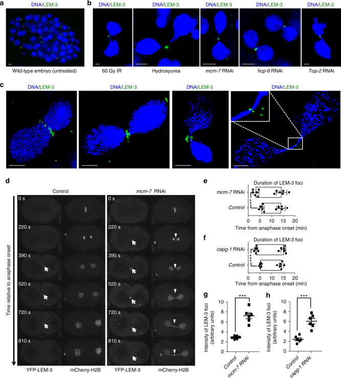
Localization of LEM-3 on chromatin bridges and at the midbody. a Localization of YFP-LEM-3 in wild-type embryo. b Localization of YFP-LEM-3 on chromatin bridges generated by IR, hydroxyurea, and RNAi depletion of replicative helicase subunit MCM-7, condensin II component HCP-6 and topoisomerase TOP-2. c Representative OMX images of YFP-LEM-3 localization at different stage of chromosome segregation in the presence of chromatin bridges induced by mcm-7 RNAi. Scale bars: 2 μm. d Localization of LEM-3 on chromatin bridges induced by mcm-7 RNAi. Arrows indicate YFP-LEM-3. Arrowheads indicate the chromatin bridges. Times are relative to anaphase onset of the first division. e, f Quantification of the average time duration of GFP-LEM-3 localization at the midzone/midbody during the first mitotic cell division in wild type, mcm-7 RNAi (e) and capg-1 RNAi (f) embryos. Error bars represent standard deviation of the mean. g, h Quantification of the intensity of GFP-LEM-3 foci during the first mitotic division in wild type, mcm-7 RNAi (g) (measured at 2.5 min after first appearance of LEM-3 foci) and capg-1 RNAi (h) embryos. Asterisks indicate statistical significance as determined by two-tailed Student t-test. p values below 0.05 were consider significant, where p < 0.05 was indicated with *, p < 0.01 with **, and p < 0.001 with ***

