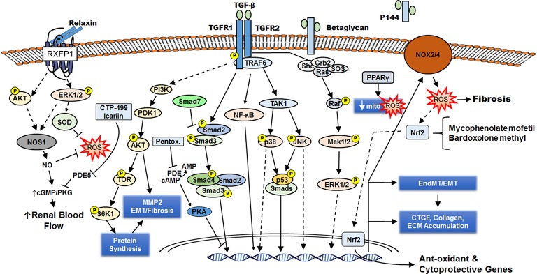
Figure 1.
Renal fibrosis occurs downstream of TGF-β intracellular signaling and increased ROS generation. Through its cell surface receptors TGFR1 and TGFR2, TGF-β activates several signaling cascades (including AKT, Smad2/3, NF-κB, p38 MAPK, JNK, ERK1/2) that lead to formation of activated fibroblasts (directly from interstitial fibroblasts or by epithelial mesenchymal transition/EMT or endothelial to mesenchymal transition/EndMT). These myofibroblasts synthesize ECM components leading to fibrosis and express NOX2/4 that generates ROS, which can enhance fibrosis or induce anti-oxidant/cytoprotective genes by activating the transcription factor Nrf2. Activation of the nuclear receptor PPARγ also reduces ROS formation by improving mitochondrial function and defenses. The cell-surface chondroitin sulfate/heparan sulfate proteoglycan betaglycan facilitates the interaction of TGF-β with TGFR2, and a soluble truncated form of betaglycan (P144) can serve as a decoy receptor to attenuate TGF-β signaling. The hormone relaxin engages the G-protein receptor RXFP1 to induce NOS1 expression and generate NO, which can directly reduce ROS or indirectly reduce ROS via increased renal blood flow. The PDE5 inhibitors CTP-499 and Icariin enhance the beneficial effects of NO on the kidney. By increasing cAMP and PKA activity, the PDE inhibitor pentoxifylline interferes with Smad3/4-dependent CTGF transcription. CTGF enhances TGF-β signaling.

