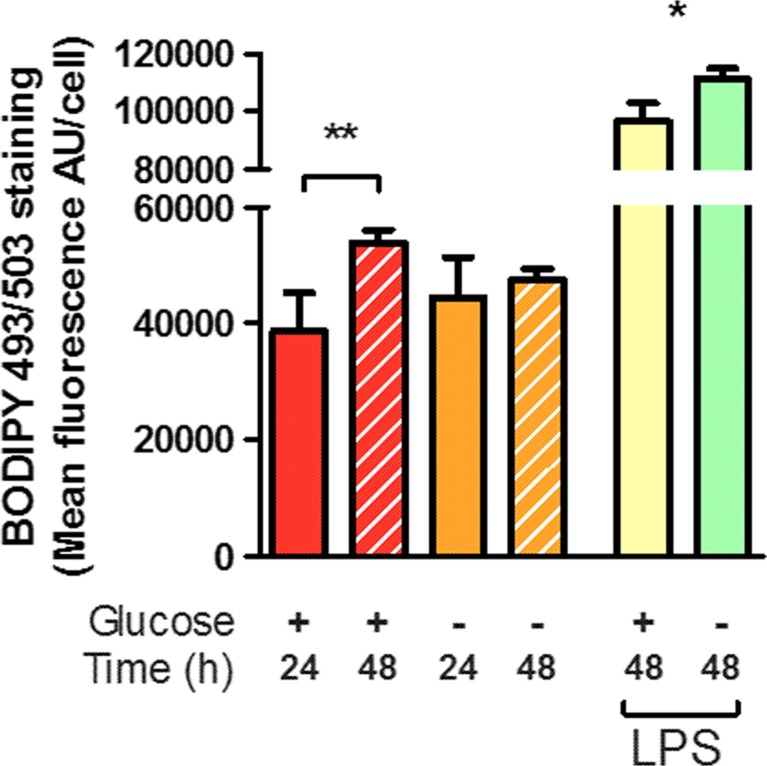
Fig. 3.
Glucose deprivation alters the accumulation of lipid droplets. Accumulation of lipid droplets was quantified by staining with BODIPY 493/503 and integration of green fluorescence within the soma of microglia identified by Hoechst 33342 and CD68 labelling, and is indicative of an increased size and/or number of lipid droplets. Fluorescence was significantly increased over the course of 48 h in the normal glucose condition, but not during glucose deprivation. Both normal and glucose-deprived cells showed significant increases in BODIPY 493/503 staining after LPS stimulation at either 24 or 48 h. Asterisk indicates significant groupwise differences by two-way ANOVA between control and LPS and Double asterisk indicates pairwise significance by Bonferroni’s post hoc (N = 4)

