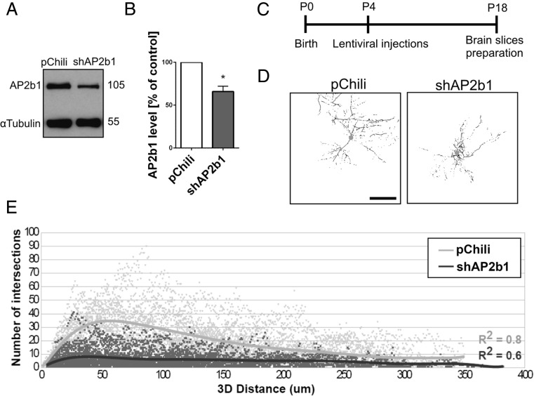
Fig. 2.
AP2 controls dendritic arbor morphology in vivo. a Western blot analysis of AP2b1 in protein lysates obtained from cultured hippocampal neurons infected with Lv-Chili or Lv-Chili-shAP2b1 on DIV4 for 6 days. b Results of quantitative WB analysis of cell lysates obtained from neurons transduced as in a. *p < 0.05 (one-sample t test). Number of independent experiments (N) = 4. Error bars indicate SEM. c The time-course of the experiment. d Representative 3D reconstructions of CA1 hippocampal neurons of rats transduced with Lv-Chili or Lv-Chili-shAP2b1 on P4 for 2 weeks. Scale bar = 100 μm. e Results of 3D Sholl analysis of CA1 hippocampal neurons from rats treated as in b. The data are expressed as the number of intersections as a function of the 3D distance from the soma. R 2 values = 0.8 for Lv-Chili (good fit) and 0.6 for Lv-Chili-shAP2b1 (satisfactory fit). Cell images were obtained from eight (Lv-Chili) or five (Lv-Chili-shAP2b1) animals. Number of cells per variant (n): Lv-Chili (28) and Lv-Chili-shAP2b1 (34)

