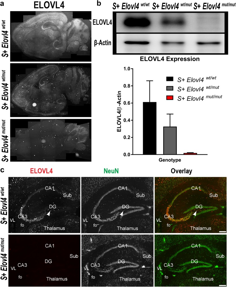
Fig. 1.
Expression of ELOVL4 in the mouse brain a ELOVL4 expression in S + Elovl4 wt/wt, S + Elovl4 wt/mut, and S + Elovl4 mut/mut mice. b Western immunoblot probing for ELOVL4 in hemisected whole brain from S + Elovl4 wt/wt, S + Elovl4 wt/mut, and S + Elovl4 mut/mut mice normalized to β-actin and quantified by densitometry. Statistics: one-way ANOVA with Tukey’s multiple comparisons test, ****p < 0.0001 (n = 6) error ± SD. c Distribution of ELOVL4 (red) co-localized with the neuronal nuclear marker NeuN (green) in the hippocampal formation in S + Elovl4 wt/wt and S + Elovl4 mut/mut mice at P20. Cornu Ammonis field 3 (CA3), polymorph layer (arrow), Cornu Ammonis field 1 (CA1), dentate gyrus (DG), subiculum (Sub), fo (fornix), VL (lateral ventricle). Scale bar = 250 μm

