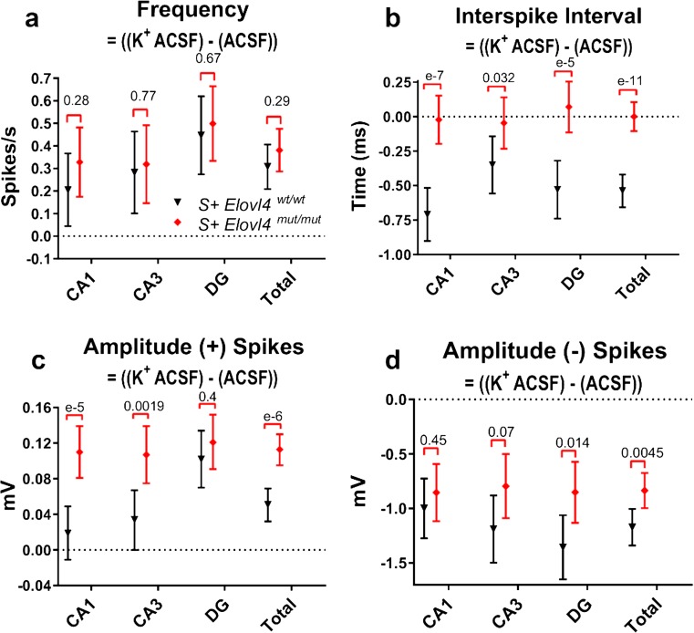
Fig. 7.
Extracellular electrophysiology under physiological conditions followed by depolarizing conditions in hippocampal slices ex vivo collected from S + Elovl4 wt/wt and S + Elovl4 mut/mut mice. The following measurements were made during 600 trace (1 s/trace) recordings of extracellular field potentials in hippocampal slices perfused with physiological ACSF (normal ACSF = 2.5 mM K+) followed immediately by a second 600 trace (1 s/trace) recording during which perfusion was switched to depolarizing, higher extracellular potassium ACSF (high K+ ACSF = 7.5 mM K+) at time = 20 s. a Evoked frequency presented as the difference between spikes/s at high K+ ACSF and spikes/s at normal K+. b Evoked inter-spike interval (ISI) presented as the time difference between spikes at high K+ and spikes at normal K+. c Evoked amplitude (+) spikes presented as the difference between spike magnitudes (mV) in high K+ and normal K+. d Evoked amplitude (−) spikes presented as the difference between spike magnitudes (mV) at high K+ and normal K+. See methods for detailed statistics (WT: n = 9, slice # = 22; mut: n = 9, slice # = 22) error ± 95% confidence interval

