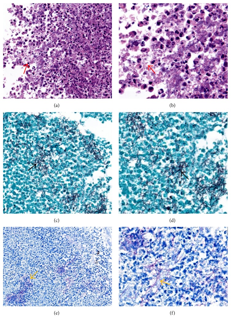
Figure 3.
Renal mass pathology shows multiple renal abscesses with filamentous structures (red arrow) on hematoxylin and eosin stain under (a) low power (40x) and (b) high power (100x). Gomori's methenamine silver stain shows no fungal hyphae but highlights the filamentous bacterial forms (black arrow) under (c) low power (40x) and (d) high power (100x). These filamentous bacteria stain weakly positive with Fite's stain supporting the diagnosis of Nocardia species (yellow arrow) under low power (40x) (e) and high power (100x) (f).

