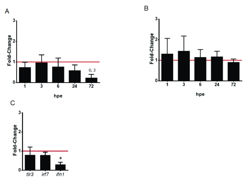
Figure 2. Interferon signaling in VHSV-exposed rainbow trout RBCs.
Time course of interferon-inducible antiviral genes mx ( A) and pkr ( B). RBCs were exposed to VHSV with a multiplicity of infection (MOI) of 1 at 14°C, and mx1-3 and pkr genes expression was quantified by RT-qPCR at time 0, 1, 3, 6, 24, 72 hours postexposure (hpe). Data is displayed as mean ± SD (n = 3). Kruskal-Wallis Test with Dunn´s Multiple Comparison post-hoc test was performed among all conditions. ( C) Interferon signaling at early time postexposure. RBCs were exposed to VHSV with a MOI of 1 at 14°C, and tlr3, irf7 and ifn1 gene expression profiles were quantified by RT-qPCR at time 0, and 3 hpe. Data is displayed mean ± SD (n = 3). Mann Whitney Test was performed for statistical analysis between the VHSV-exposed and control cells (non-exposed, time 0, red line). Gene expression was normalized against eukaryotic 18S rRNA for mx, tlr3, irf7 and ifn1 genes and ef1α for pkr gene, and relativized to control cells (fold-change). Asterisk denote statistically significant differences between VHSV-exposed and control cells ( P-value < 0.05).

