Figure 3.

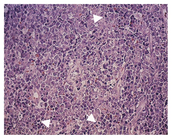
Histological aspect of the specimen revealing a diffuse proliferation of atypical large lymphoid cells, with predominance of centroblasts and high mitotic index (arrows) (HE ×100).

Histological aspect of the specimen revealing a diffuse proliferation of atypical large lymphoid cells, with predominance of centroblasts and high mitotic index (arrows) (HE ×100).