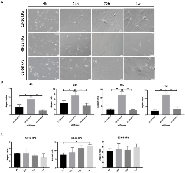
Figure 2.
Morphology of BMMSCs on gels with various stiffnesses. (A) After BMMSCs were planted on the gels, the cells were analyzed with an inverted phase contrast microscope at 4h-1w. Scale bar = 20 μm. (B, C) Quantification of morphological changes versus stiffnesses at 4 h, 24 h, 72 h and 1w. Cell aspect ratio was measured. * P < 0.05, ** P<0.01.

