Figure 9.
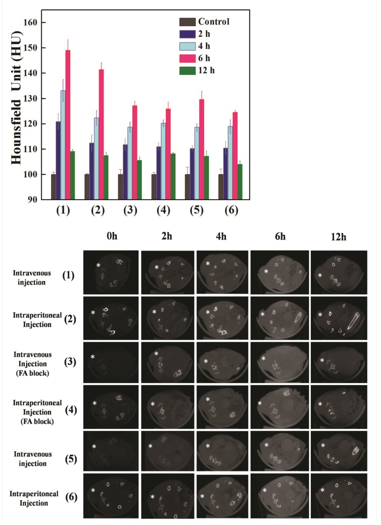
Representative transectional micro-CT images (a) and CT signal intensities (b) of the HeLa cells xenografts in nude mice before (0 h) and 2, 4, 6 and 12 h after exposure to [(Au0)300-G5.NHAc-(PEG-FA)-mPEG] DENPs 1-4 and [(Au0)300-G5.NHAc-mPEG] DENPs (5 and 6) via different admission routes. The tumor areas were labeled with “*”. 1 × 10⁶ HeLa cells were subcutaneously injected on the right side of the backs of 4-to 6-week-old male BALB/c nude mice. Approximately 3 weeks after the injection, the mice were then placed in a scanning holder and scanned with a micro-CT imaging system as described in the Materials and Methods. The data shows that treatment with Au DENPs-FI-FA-DTA increases the CT signal intensities in the HeLa cell xenografts, which can be blocked by pretreatment with free FA. These results further demonstrate the roles of the FA moieties in mediating the specific targeting of our nanoparticles. (300 × 300 DPI).

