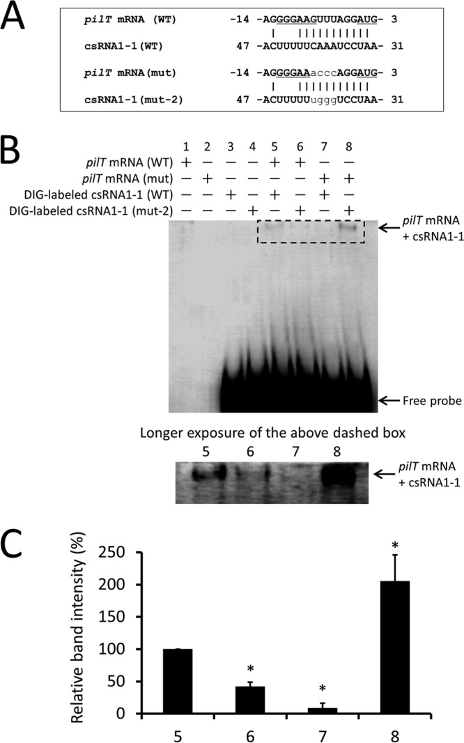
FIG 5.

RNA-RNA electrophoretic mobility shift assay of csRNA1-1 with pilT mRNA. (A) Putative interactions between the 5′ end of pilT mRNA (WT) and csRNA1-1 (WT) or pilT mRNA (mut) and csRNA1-1 (mut-2). The ribosome binding site and start codon are underlined. Substituted nucleotides in mutants are shown in plain lowercase. (B) DIG-labeled csRNA1-1 WT or csRNA1-1 mut-2 was incubated with pilT mRNA (WT) or pilT mRNA mutant (mut). Reaction products were separated on 5% native polyacrylamide gel and transferred to a membrane. DIG-labeled RNA signals on the membrane were detected using anti-DIG antibody. The image shown is representative of three independent experiments. The shifted bands in lanes 5 to 8 (the region surrounded by a dashed-line rectangle) are shown with a longer exposure time in the lower panel. (C) Quantification of the intensity of shifted bands in lanes 5 to 8. Data are expressed as means ± SD from three independent experiments. *, P < 0.01.
