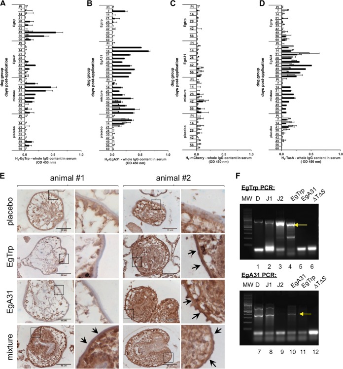
FIG 4.
A specific humoral response is elicited in dogs after oral application of recombinant B. subtilis spores harboring Echinococcus granulosus antigens. Dog serum was collected on the indicated days postapplication and tested by indirect ELISA using plates coated with E. coli purified H6-EgTrp (A), H6-EgA31 (B), H6-mCherry (C), and H6-TasA (D). The samples were then incubated with specific dog anti-IgG conjugated with HRP. The dog groups are indicated on the vertical axis. White and black bars, dogs 1 and 2 in each group, respectively. The data represent the mean ± SEM from three independent experiments. (E) Immunohistochemistry of sheep E. granulosus protoscoleces incubated with the indicated dogs sera (1:100) followed by secondary anti-IgG dog-HRP and stained with diaminobenzidine (brown). Nuclei were counterstained with hematoxylin (blue). Each row corresponds to the indicated dog group (groups treated with placebo, EgTrp, EgA31, and the mixture of EgTrp and EgA31). The number of the dog to which the treatment was orally applied is reported at the top of each panel. The right columns of the two panels correspond to enlargements of the insets marked with a black frame in the pictures on the left. Black arrows, positive sera recognizing the subtegument/tegument membrane of the protoscoleces. Bars, 50 μm. (F) PCR characterization of intestinal recombinant B. subtilis bacteria isolated from the small intestine of dog 2 from the mixture group. Colonies isolated from one duodenal section (lanes D) and two jejunal sections (lanes J1 and J2) were analyzed by PCR for the detection of the B. subtilis tasA sinR TasA-EgTrp (top) and B. subtilis tasA sinR TasA-EgA31 (bottom) strains. Each panel also includes the results for a set of controls, namely, EgTrp, EgA31, and ΔTΔS, which correspond to laboratory strains B. subtilis tasA sinR TasA-EgTrp, B. subtilis tasA sinR TasA-EgA31, and B. subtilis tasA sinR, respectively. Lanes MW, molecular weight markers; yellow arrows, the position of each E. granulosus-specific band.

