FIG 4.
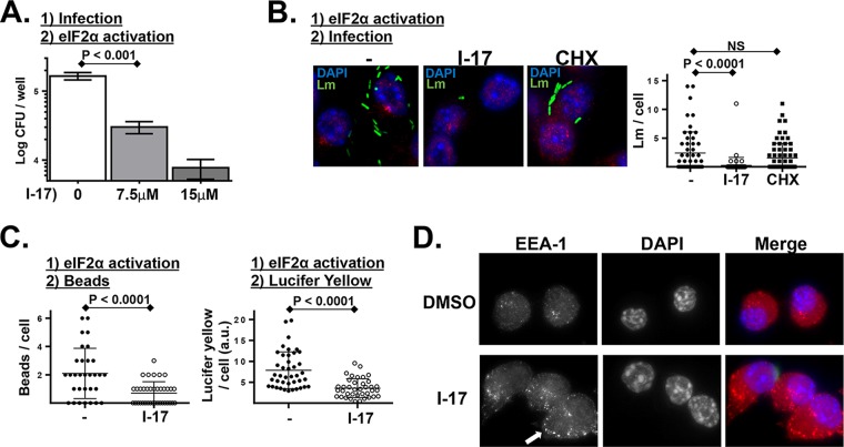
L. monocytogenes infection is inhibited in cells treated with a small-molecule activator of eIF2α phosphorylation. (A) PEMs were infected with L. monocytogenes for 30 min, noninternalized bacteria were killed with gentamicin, and then cells were treated with the indicated concentration of eIF2α phosphorylation activator I-17. Following 6 h of infection, intracellular L. monocytogenes levels were determined by CFU assay. (P values were calculated using a Student t test of results of a single representative experiment performed three times.) (B) Cultured murine macrophage-like cells (RAW 267.4) were left untreated or treated with I-17 or treated with cycloheximide (CHX) for 60 min. Cells were then infected with GFP-expressing L. monocytogenes (Lm) for 45 min, washed extensively, and analyzed by fluorescence microscopy. Plotted data represent the number of L. monocytogenes bacteria per cell from a representative experiment performed multiple times with similar results. NS, not significant. (C) Similar experiment in which untreated and I-17-treated macrophages were pulsed with fluorescent beads (left plot) for 5 min or with the soluble dye Lucifer yellow (LY) for 20 min (right plot), washed extensively, and then analyzed by fluorescence microscopy. Plotted data represent the number of beads or level of fluorescence per cell from a representative experiment performed three times with similar results. a.u., arbitrary units. (D) Similar experiment in which cells were treated with either vehicle alone or I-17 (10 μM) for 60 min and then were fixed and stained for the early endosomal marker EEA1. The arrow indicates the cumulative early EEA1-positive endosomes in I-17-treated cells.

