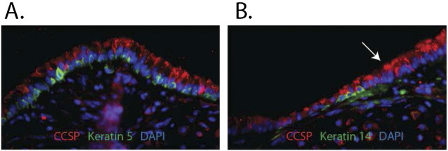
Figure 1.
Mouse tracheal histology. Tracheal sections from normal mice were stained using dual immunofluorescence methods. (A) The intercartilaginous region. Clara-like cells are detected as CCSP+ (red) cells. Basal cells are detected as Keratin (K) 5+ (green) cells. Note the pseudostratification in this region of the trachea. (B) The midcartilaginous region. Clara cells are stained as indicated above. The K14+ subset of basal cells is shown by K14 (green) staining. Note that this region is less stratified than the intercartilaginous region. Arrow indicates the transitional zone between the two regions.

