
Figure 5.
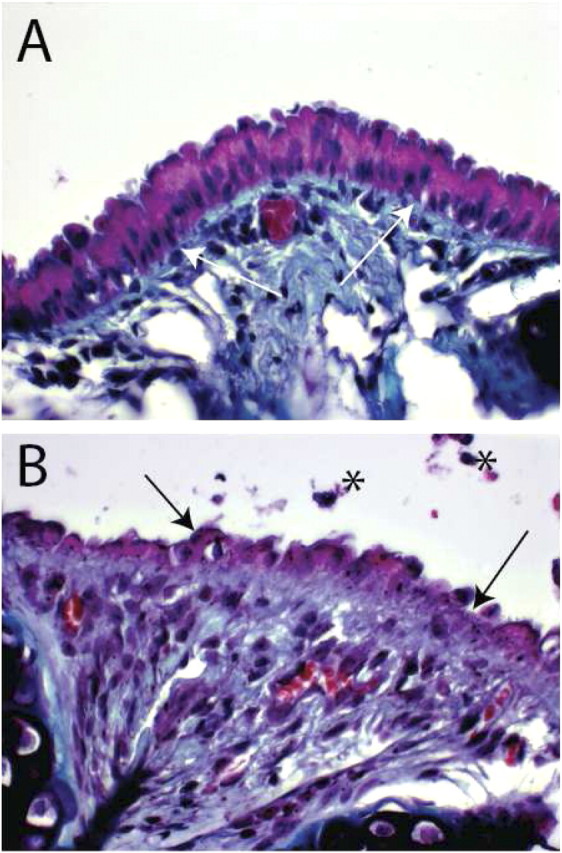
Tracheal histology. (A) An intercartilaginous region from a normal mouse. Periodic Acid Schiff stain. White arrows indicate the basement membrane. (B) An intercartilaginous region from a mouse that was treated with naphthalene and recovered for 2 days. Black arrows indicate the disrupted epithelium. Note that the boundary between the injured epithelium and the subepithelial space is distorted and that the subepithelial compartment is hyperplastic. Asterisks indicate cellular debris.