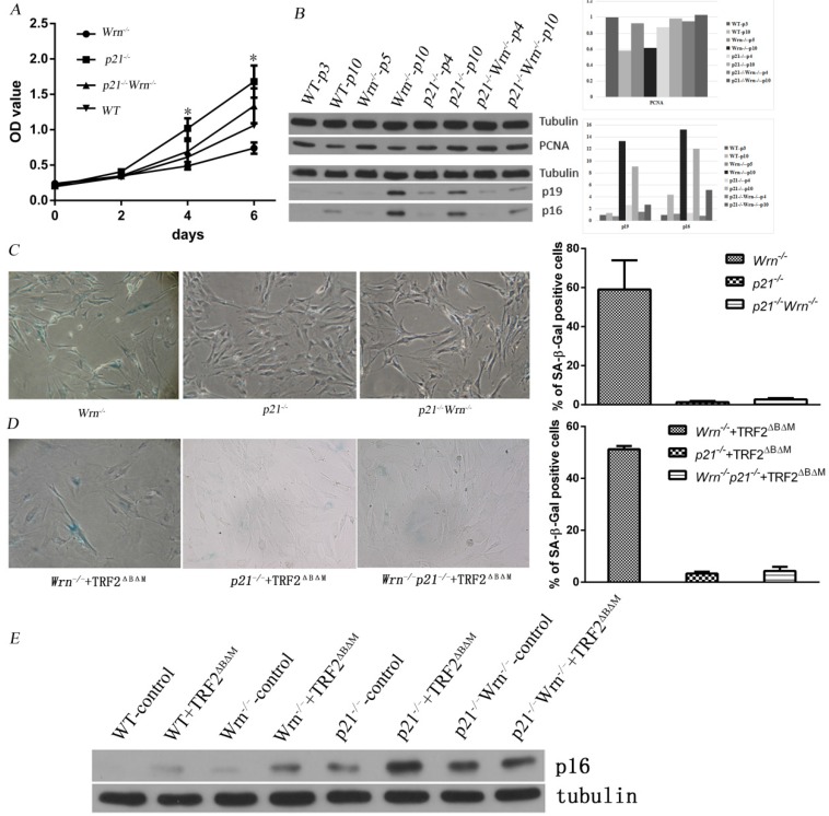
Figure 1.
Loss of p21 function rescued cellular senescence induced by telomere dysfunction. A. Growth curves of WT, Wrn-/-, p21-/- and p21-/-Wrn-/- MEFs. The asterisk in day 4 showed the statistical significant difference between Wrn-/- and p21-/- MEFs (p= 0.02). The asterisk in day 6 showed the statistical significant difference between Wrn-/- and p21-/- MEFs (p= 0.01), and between Wrn-/- and p21-/-Wrn-/- MEFs (p=0.02). B. Analysis of PCNA, p16 and p19ARF (p19) expression levels in MEFs with different genotypes in their early passages (passage number≤5) or late passages (passage number=10) by Western blot. The quantification result was showed at the right side. C. SA-β-Gal staining of p21-/-, Wrn-/- and p21-/-Wrn-/- MEFs in late passage (passage number=13), showing that loss of p21 function could rescue the cellular senescence induced by Wrn-/-. The quantification result was showed at the right side. D. SA-β-Gal staining of p21-/-+TRF2ΔBΔM, Wrn-/+TRF2ΔBΔM and p21-/-Wrn-/-+TRF2ΔBΔM MEFs, showing that even with telomere dysfunction caused by both Wrn and TRF2 dysfunction, loss of p21 function could still rescue the cellular senescence. The quantification result was showed at the right side. E. Compared to the control WT MEFs, MEFs with p21 knocking out showed higher level of p16 expression.

