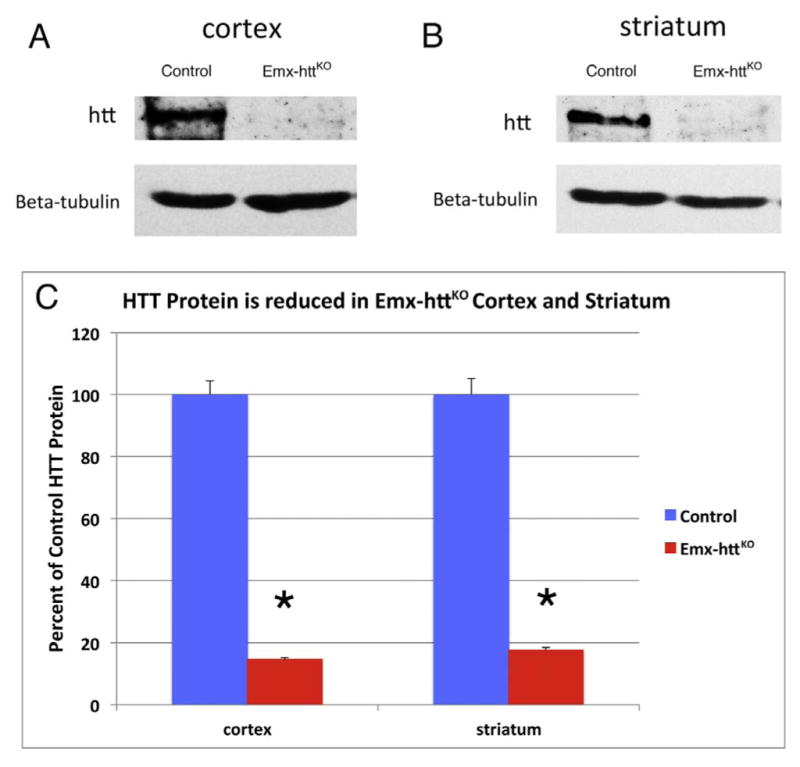
Fig. 2.

Western blot analyses of total protein lysates from cortex and striatum of control and Emx-httKO mice. Protein lysates were separated in 8% SDS-PAGE and transferred to nitrocellulose membrane. The upper panel in image A shows detection of huntingtin with a mouse monoclonal anti-huntingtin antibody 2166, and the lower panel shows anti-β-tubulin staining as a loading control, from 5-month-old control and Emx-httKO mice. Note that huntingtin protein is substantially reduced in cortex of Emx-httKO mice, confirming knockout efficacy. Huntingtin protein was also substantially reduced in striatum, as shown in image B comparing 5-month-old control and Emx-httKO mice. As explained in the text, this is likely to largely represent depletion of huntingtin from corticostriatal terminals, which appears to be the predominant source of huntingtin in striatum. The graph in image C shows densitometric analysis of Western blot results for huntingtin in cerebral cortex and striatum in 5 control and 5 Emx-httKO mice at 5- or 12-months of age, normalized to tubulin, confirming that the reduction of huntingtin in Emx-httKO cortex and striatum is highly significant (asterisks).
