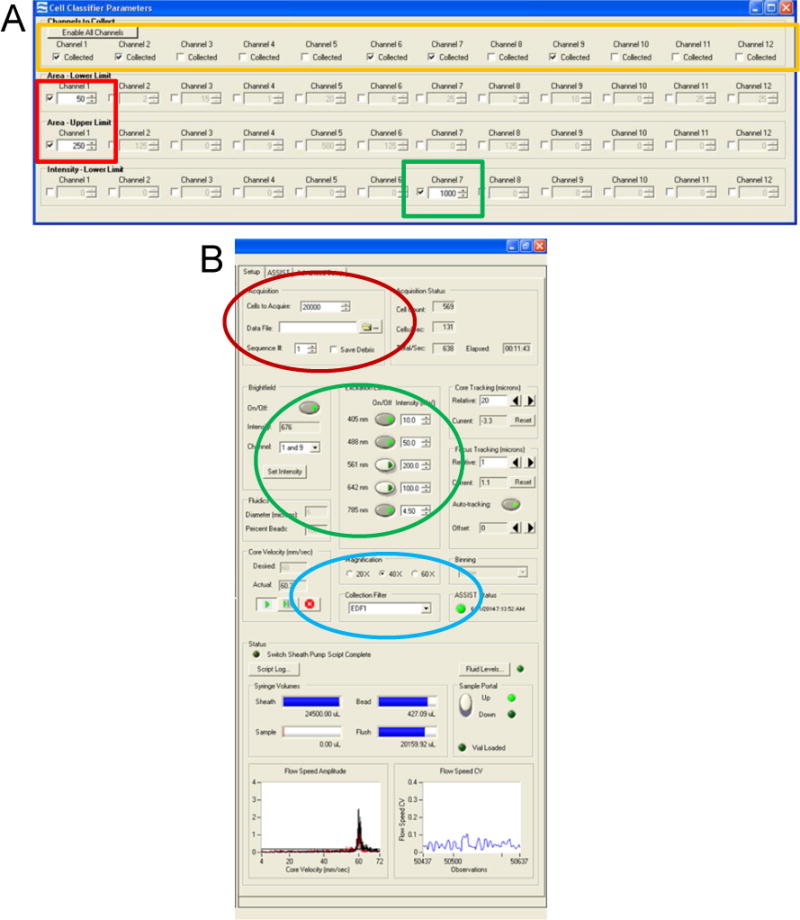
Figure 1. Setting up the Inspire software.

(A) Cell Classifiers panel with the Area Lower Limit and Area Upper Limit for channel 1 brightfield selected (red box). Intensity Lower Limit of channel 7 is selected to collect events with a positive nuclear signal (green box), and the desired channels selected (yellow box). (B) Experimental set-up panel on the left of the user interface is highlighted. Acquisition parameters (red oval), illumination (green oval), and magnification with EDF filter (blue) are shown.
