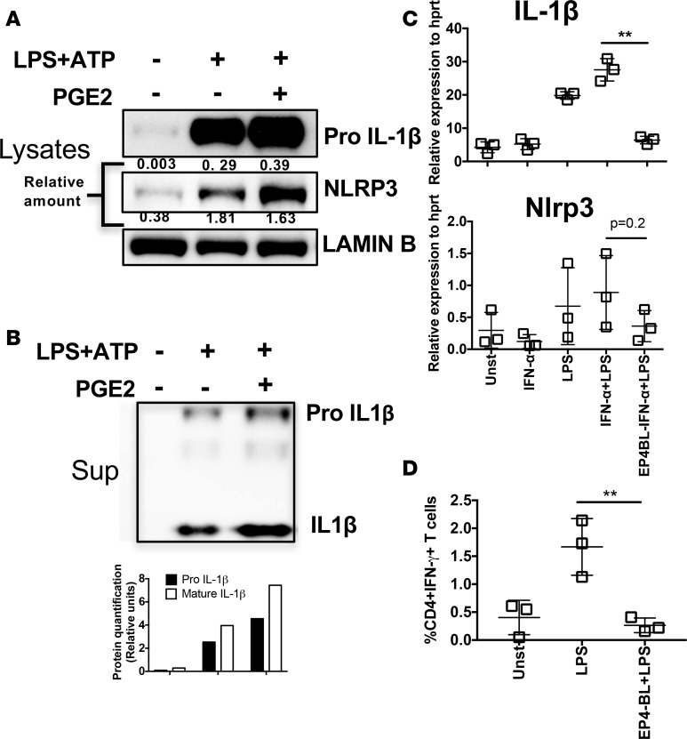
Figure 5. PGE2-PTGER4–driven signals are inflammatory in NOD mice.
(A and B) Spleen cells from 8-week-old NOD mice were stimulated with LPS-ATP. After 4 hours of LPS stimulation, where indicated PGE2 was added to the culture for 30 minutes, followed by ATP stimulation for 1 hour. Cells lysates (A) or supernatant (B) were analyzed by Western blotting for IL-1β and NLRP3. Protein bands were quantified relative to LAMIN-B (A) or total cell number (B). (C) CD11c+ cells were enriched and stimulated in vitro with IFN-α or LPS or both and PTGER4 (EP4) antagonist (EP4BL) as indicated. Transcripts of IL-1β and Nlrp3 were measured 2 hours after stimulation. (D) Total spleen cells were stimulated with or without LPS with or without PTGER4 antagonist. Intracellular IFN-γ was measured 6 hours after stimulation in CD4+ T cells. Data are representative of at least 2 (A and B) or 3 (C and D) independent experiments performed with pooled mice (n = 3), mean ± SD. Statistical analysis was performed with 1-way ANOVA with Bonferroni post-test, and only the relevant comparison was depicted (C and D). **P < 0.01.

