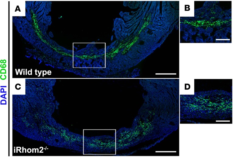
Figure 2. Localization of CD68+ immune cells within the infarct region of iRhom2-deficient (iRhom2–/–) hearts at day 7 after myocardial infarction (MI).

Seven days after MI, (A and B) wild-type hearts revealed CD68+ myeloid cells (green) densely localized and highly organized within the infarct region. However, (C and D) iRhom2 mutants had an expression pattern of CD68+ immune cells that were more dispersed throughout the infarct zone. Representative images are based on 3 biological replicates. Scale bars: 500 μm (A and C), 200 μm (B and D).
