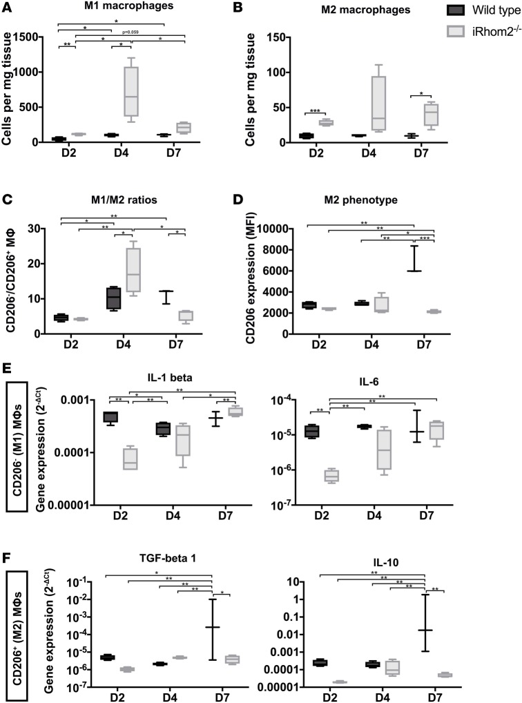
Figure 6. M1/M2 macrophage ratios and cytokine expression within the first week after injury.
The numbers of (A) CD206– M1 macrophages (MΦs) were significantly increased at days 2 and 4 in iRhom2-deficient (iRhom2–/–) hearts compared with controls. Similarly, the numbers of (B) CD206+ M2 MΦs were increased significantly at days 2 and day 7 in iRhom2–/– hearts. (C) M1/M2 ratios were increased and decreased at days 4 and 7, respectively, in iRhom2–/– mutants compared with wild type. (D) The mean fluorescence intensity (MFI) of CD206+ M2 MΦs was markedly reduced in iRhom2–/– MΦs at day 7 after injury. (E) Relative mRNA expression (2–ΔCT) of proinflammatory cytokines (Il1b and Il6) from sorted M1 MΦs (CD45+CD64+MerTK+Ly6G–F4/80+CD206–) was significantly decreased at day 2 after injury compared with wild-type controls. (F) Gene expression of reparative cytokines (Il10 and Tgfb1) were significantly reduced in sorted M2 MΦs (CD45+CD64+MerTK+Ly6G–F4/80+CD206+) at day 7 after injury, compared with wild-type hearts. Data are shown as the mean ± SEM, n = 5 for each experimental group. *P ≤ 0.05, **P ≤ 0.01, ***P ≤ 0.001, 1-way ANOVA and post-hoc test.

