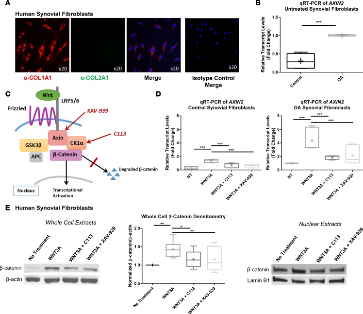
Figure 2. Synovial fibroblasts are responsive to Wnt inhibition treatment.
(A) Synovial fibroblasts isolated from patients with osteoarthritis (OA) were positive for expression of type I collagen (red) but not type II collagen (green). Nuclei were stained with DAPI (blue). Isotype controls were negative. Original magnification, ×20. (B) OA synovial fibroblasts were compared with control synovial fibroblasts by real-time qRT-PCR of AXIN2, a downstream marker of Wnt signaling. Expression was higher in synovial fibroblasts isolated from patients with OA than those from controls. Statistical analysis was performed using a t test, ***P < 0.001, n = 6. (C) Schematic of the Wnt/β-catenin pathway showing target molecules inhibited by the Wnt inhibitors (C113 and XAV-939) used in the study. (D) Furthermore, synovial fibroblasts were treated with recombinant WNT3A with and without Wnt inhibitors (C113 and XAV-939) and analyzed by real-time RT-PCR of AXIN2. Wnt signaling was ameliorated by both inhibitors in control and OA synovial fibroblasts. One-way ANOVA with Tukey’s post-hoc test was used to compare treatment groups, ***P < 0.001, n = 6. (E) Immunoblotting of β-catenin in OA synovial fibroblast cell lysates after treatment with recombinant WNT3A with or without inhibitors. Densitometry of immunoblots was performed to quantify the reduction of Wnt signaling after inhibitor treatment. One-way ANOVA with Tukey’s post-hoc test was used to compare treatment groups, *P < 0.05, **P < 0.01, n = 8. Alteration of β-catenin signaling in the nucleus was confirmed by immunoblotting of synovial fibroblast nuclear lysates compared with lamin B1 loading control. (B, D, and E) For data presented as box-and-whiskers plots, horizontal lines indicate the medians, cross marks indicate the means, boxes indicate the 25th to 75th percentiles, and whiskers indicate the minimum and maximum values of the data set.

