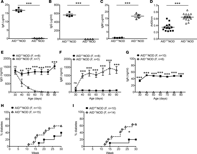
Figure 2. Early exposure to maternal IgG is dispensable for T1D development in AID–/–NOD mice.
(A–C) Serum Igs from 2-month-old nondiabetic female AID–/–NOD mice and AID+/+NOD littermates were measured by ELISA. (A) IgA; (B) IgG; (C) IgM. Data are shown as mean ± SEM from 1 of at least 2 independent experiments. n = 4 mice/group. (D) Total anti-insulin Ig (IgH+L) measured by ELISA using sera from 2-month-old nondiabetic female AID–/–NOD mice and AID+/+NOD littermates. Data are presented as OD of 405 nm and shown as mean ± SEM by pooling 3 independent experiments. n = 13–14 mice/group. (E–G) Dynamic changes in serum Igs. Sera were taken from 30-day-old female AID–/– and AID+/– NOD mice and then every 10 days thereafter followed by Ig measurement by ELISA. Data are shown as mean ± SEM and were pooled from 2 independent experiments. n ≥ 5 mice/group. (H) Diabetes incidence in female progeny of female AID+/–NOD and male AID–/–NOD breeding. (I) Diabetes incidence of female progeny of female AID–/–NOD and male AID+/–NOD breeding. Data were pooled from 2 independent experiments. n ≥ 10 mice/group. *P < 0.05; **P < 0.01; ***P < 0.001, Student’s t test (A–G) and Gehan-Breslow-Wilcoxon survival test (H and I).

