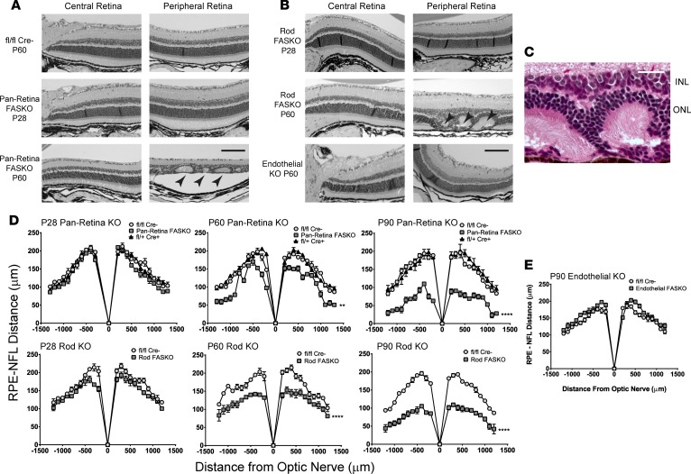
Figure 2. Loss of FAS activity is associated with progressive retinal degeneration.
(A) H&E-stained paraffin cross sections of neural retinal layers, with pseudorosette formation (arrowheads) in P60 FASKO compared with P28 FASKO and to controls. (B) Images from rod-specific FASKO at P60 compared with P28, as well as normal images from endothelial FASKO mice. (C) Representative detail of outer retinal pseudorosettes seen in both rod FASKO and pan retinal FASKO, with radially arranged and apposed photoreceptor outer segments. INL, inner nuclear layer; ONL, outer nuclear layer. (D and E) Quantification of retinal thickness changes in neural and endothelial FASKO compared with littermate controls (n = 4 animals/group for P28, P60, and P90 Pan Retina KO, P90 Rod KO, and P90 Endothelial KO panels; n = 6 animals/group for P28 and P60 Rod KO panels; 2-way ANOVA). Scale bar: 100 μm (A and B) and 25 μm (C). **P < 0.01, ****P < 0.0001.

