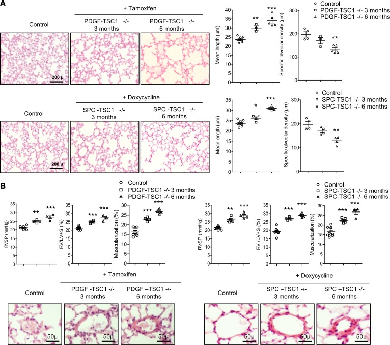
Figure 8. Effects of mTOR overactivation in SPC-TSC1–/– and PDGF-TSC1–/– mice.
TSC1 deletion was induced by i.p. tamoxifen in PDGF-TSC1–/– mice and by treatment with doxycycline in drinking water in SPC-TSC1–/– mice. The mice were investigated 3 and 6 months later and were compared with vehicle-treated mice. (A) Development of lung emphysema in PDGF-TSC1–/– and SPC-TSC1–/– mice. From left to right, representative lung sections stained with H&E and showing emphysema lesions in PDGF-TSC1–/– and SPC-TSC1–/– mice, mean linear intercept of alveolar septa, and air space. Scale bars: 200 μm. (B) Development of pulmonary hypertension in PDGF-TSC1–/– and SPC-TSC1–/– mice. Right ventricular systolic pressure (RVSP), right ventricular/left ventricular + septum weight ratio (RV/LV+S), and muscularization of pulmonary vessels (percent of muscularized vessels over the total number of pulmonary vessels). Representative pulmonary vessels in PDGF-TSC1–/– and SPC-TSC1–/– mice compared with control mice. *P < 0.05, **P < 0.01, ***P < 0.001 vs. control mice (1-way ANOVA). Scale bars: 50 μm.

