Figure 3.
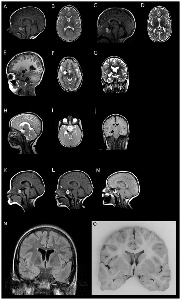
Representative brain MRI findings in individuals with PURA syndrome. Subject DB13-043, a 5 year old girl with hypotonia and epilepsy, had thinning of the corpus callosum (A) and of the subcortical white matter with increased extra-axial fluid spaces (B). Subject DB15-027, a 4 year old boy with hypotonia and cortical visual impairment, had mildly increased extra-axial fluid in the posterior fossa (C) and thinning of the cerebral white matter (D). Subjects DB15-021 (E–G) and DB15-033 (H–J) had subarachnoid cysts (arrowhead). Subject DB16-002 had thickening of the corpus callosum and cerebellar tonsilar ectopia (asterix) develop over scans at ages 2 years (K), 6 years (L), and 9 years (M). Coronal T2 FLAIR image from subject DB16-016 at age 20 years showing mild dilation of the lateral ventricles and increased signal in the subcortical white matter (N). Gross brain specimen of subject DB16-016 confirming ventricular and white matter findings seen on neuroimaging (O).

