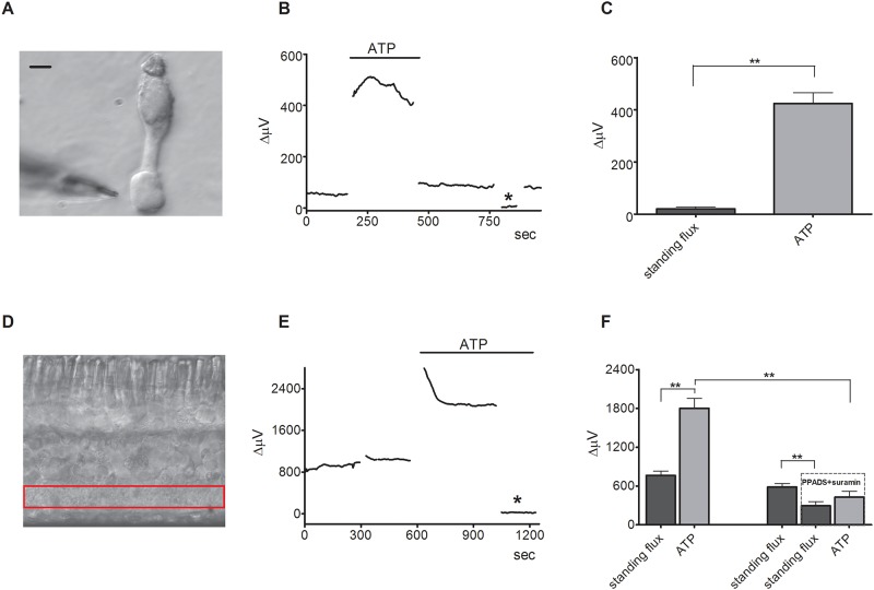
Fig 6. Extracellular ATP also induces increases in extracellular H+ fluxes from the end feet of isolated Müller cells and the inner plexiform layer in retinal slices that are reduced by PPADS and suramin.
(A) An isolated Müller cell with an H+-selective microelectrode positioned by the end foot of the cell; scale bar, 20 μm. (B) A representative trace showing the increase in H+ flux measured from the end foot of a Müller cell in response to application of 100 μM ATP. (C) Mean responses of isolated Müller cells to 100 μM ATP; N = 9, asterisk denotes background control taken 200 μm above the cell. (D) Retinal slice with red box highlighting the inner plexiform layer (IPL). (E) A representative trace showing the response observed to application of 100 μM ATP from a self-referencing H+-selective microelectrode from a retinal slice when the microelectrode was positioned just above the IPL; asterisk indicates a background control reading taken 600 μm above the retinal slice. (F) Mean data from two populations; ATP induced a significant increase in H+ flux that was significantly smaller in the presence of suramin and PPADS (N = 6) than it did in normal Ringer’s solution (N = 6). The data from the two populations are separated by the gap on the x-axis and were compared via an unpaired 2-way t test.

