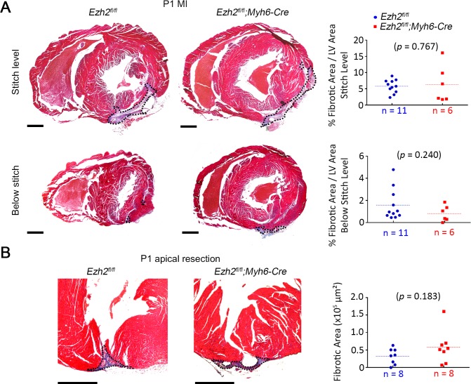
Fig 3. Ezh2 is not required for innate neonatal cardiac regeneration.
(A) Masson’s trichrome-stained cross sections showing fibrous tissue at the level of the ligature (stitch) and below the ligature in control (Ezh2fl/fl) and mutant (Ezh2fl/f;Myh6-Cre) hearts, 3 weeks post-MI induced at P1. Scar area is outlined. Plots represent quantification of fibrotic scar area as the percentage of the left ventricle (LV) area. (B) Masson’s trichrome-stained sections showing fibrous tissue (blue) at the resection area in control (Ezh2fl/fl) and mutant (Ezh2fl/fl::Myh6cre+) hearts 3 weeks post apical resection at P1. Plots represent quantification of the scar area. Scar area is outlined. Dotted lines show the mean in plots. Scale bar = 500μm.

