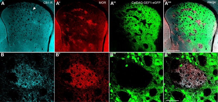
Fig 4. CB1-R immunoreactivity is enriched in striatal striosomes.
A coronal hemisection through the left striatum shows immunofluorescence for CB1-R (A, B), striosomal MOR (A’, B’) and direct eGFP fluorescence in the matrix (A”, B”) in a CalDAG-GEFI-eGFP mouse, imaged by confocal microscopy. Merged images (A”‘, B”‘). The arrow in (A) designates the striosome shown in a magnified view (B-B”‘). Images were taken with the FV 1200 confocal microscope using the MIT immunohistochemistry protocol. Scale bar is 500 μm in A”‘, 50 μm in B”‘.

