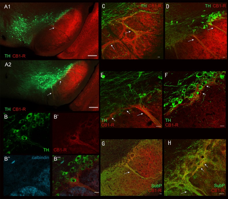
Fig 6. Lateral localization of CB1-R-dense striosome-dendron bouquets and ventral tier SNpc enveloping CB1-R-immunoreactive axons through the substantia nigra.
Two low-power images (A1, A2) show apparent dendron structures and their location. Calbindin (blue) labels the fibers in the SNpr (B”) and avoids the SNpc and dendron (labelled by TH; B, B”‘). CB1-R labels the ventrally extending dendron (B’, B”‘). An example of a TH- and CB1-R-positive dendron in the rostral SN is shown in C. Low (D) and higher power images (E, F) of the lateral SN show a CB1-R-labelled dendron extend medially into the adjacent sector of SNc (arrows in E, F). A low-power (G) and confocal image (H) of CB1-R (red) and Substance P (green) shows overlap in the dendron bouquets. Scale bars are 20 μm in B-H. Low-power images were taken with the Axiozoom. Confocal images were taken with the LSM510.

