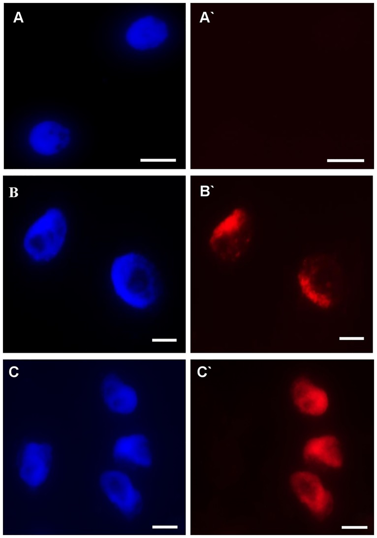
Fig 12. Examples of the detection of S-phase cells using EdU incorporation in roots of barley cv. ‘Sebastian’ seedlings treated with different concentrations of Al for 1 and 7 days.
(A-C) DAPI staining (A`-C`) Alexa Fluor 647 fluorescence (red) in S-phase cells detected after EdU incorporation. Bars represent 20 μm.

