
Fig 4. Functional GFP-tagged 19S RP subunits consist of a prominent nuclear and a smaller cytoplasmic subpopulation.
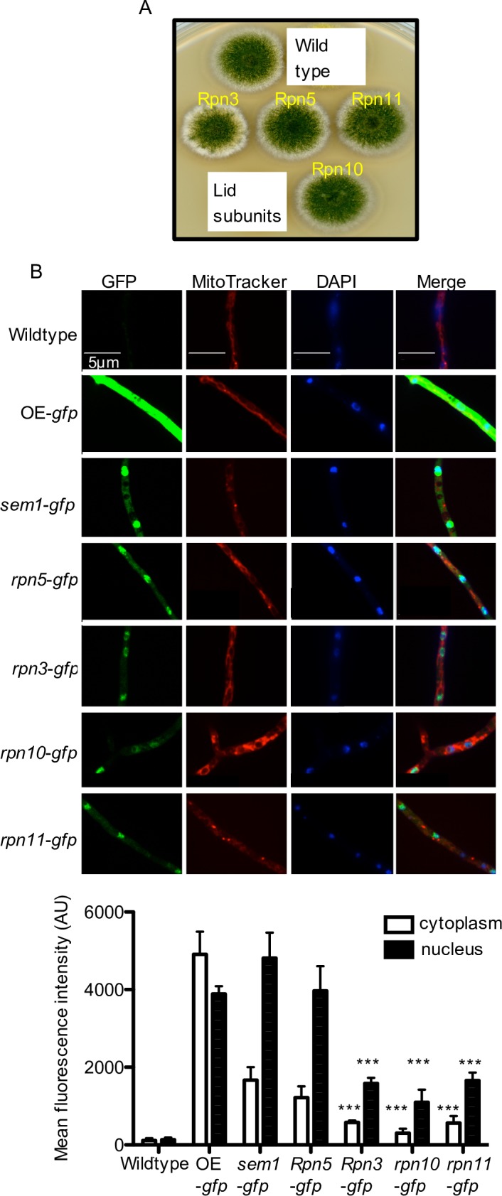
(A) Indicated 19S RP subunits fused to GFP are functional and support asexual development. (B) Subcellular localization of GFP-tagged 19S RP strains determined by fluorescence microscopy. OE-overexprssion strain. The images show green GFP fluorescence (left) for 19S RP subunits, MitoTracker Red (second from left) for mitochondria, DAPI for nuclei (third from the left), and an overlay (most right). The fluorescence intensities in the cytoplasm and nucleus for the indicated GFP strains are shown as mean fluorescence intensity. Significance of differences was calculated with t-test compared to sem1-gfp, ***p<0.001.