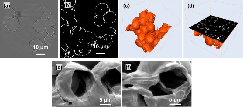
Figure 7.
(a) Transversal virtual cut of PCL microspheres showing XCT phase contrast based fringes. (b) Transversal virtual cut of PCL microspheres after shell segmentation using phase contrast based fringes. (c) 3D visualization of segmented hollow PCL microspheres. (d) 3D visualization of segmented hollow spheres and the corresponding orthogonal virtual cut. (e, f) SEM images showing the wall between merged microspheres.

