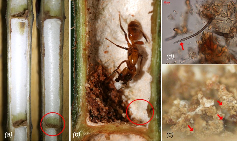
Fig 1. Colonization of a Cecropia sp. domatium.
(a) Parenchyma (white tissue) of the inner domatium wall. The part from where it has been scraped off is marked with a circle; (b) An Azteca xanthochroa queen with a parenchyma pile inoculated with chaetothyrialean fungi (foundress patch). Eggs and larvae are deposited next to the fungal patch (circle); (c) Detail of an Azteca constructor foundress patch with hyphae and (d) conidiophores (arrowheads). Scale bars: (a) 2cm, (b) 2mm.

