
Fig. 1.
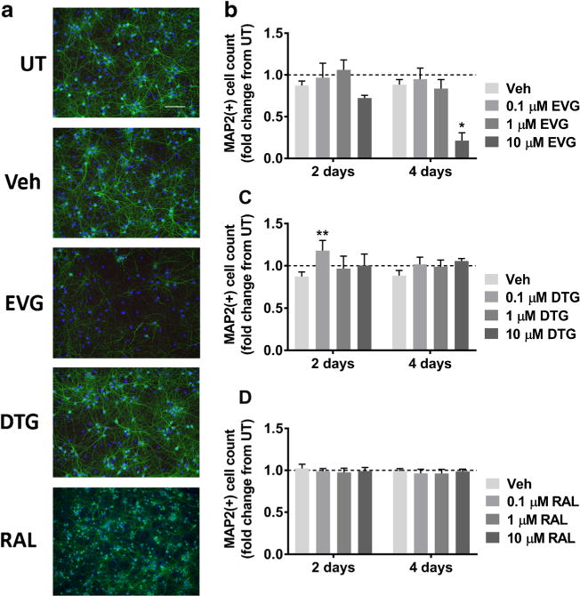
EVG but not DTG or RAL is toxic to primary rat cortical neuroglial cultures. a Cultures were treated with DMSO vehicle or 0.1 μM, 1 μM, or 10 μM EVG, DTG, or RAL for either 2 days or every other day for 4 days. Representative images of neuroglial cultures immunostained for MAP2 (green) and DAPI (blue) after treatment with 10 μM of indicated compounds for 4 days are shown at 20× magnification. Scale bar represents 100 μM. b-d Quantification of MAP2+ cells treated with indicated compounds is shown (repeated measures two-way ANOVA followed by Dunnett’s test, n = 3, *p < 0.05, **p < 0.01 vs drug vehicle). Dashed lines represent untreated (UT) cultures