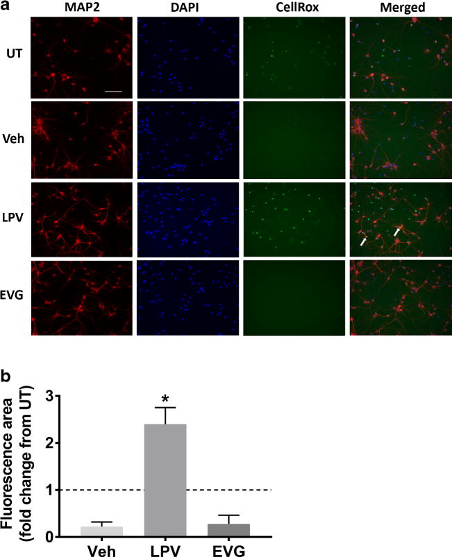
Fig. 3.

LPV but not EVG induces oxidative stress. a Rat cortical neuroglial cultures were treated with DMSO vehicle or 10 μM LPV or EVG for 1 h prior to the addition of CellRox Green reagent and live cell imaging. Images captured by time-lapse live imaging were merged with the images of the same cells that were subsequently fixed and immunostained for MAP2 and DAPI. Representative images captured 30 min following CellRox addition show cells immunostained for MAP2 (red), DAPI (blue), and CellRox green at 20× magnification. Scale bar represents 100 μm; white arrows indicate examples of neurons that accumulated CellRox green dye. b Quantification of the area positive for CellRox green fluorescence normalized to untreated (UT) cultures (dashed line) is shown (repeated measures one-way ANOVA followed by Dunnett’s test, n = 4, *p < 0.05 vs drug vehicle)
