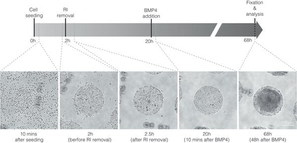
Figure 1. Timeline of the morphology of micropatterned colonies.

The images show the morphology of the micropatterned hESC colonies under phase microscopy 10 minutes after seeding (Step 11), before removal of ROCK inhibitor (Step 13), after removal of ROCK inhibitor (Step 14), 10 minutes after BMP4 addition (Step 16), and 48 hours after BMP4 addition (Step 17). RI=ROCK inhibitor. ESCRO institutional regulatory board permission was obtained to perform these experiments. Scale bar = 200 μm, applies to all panels.
