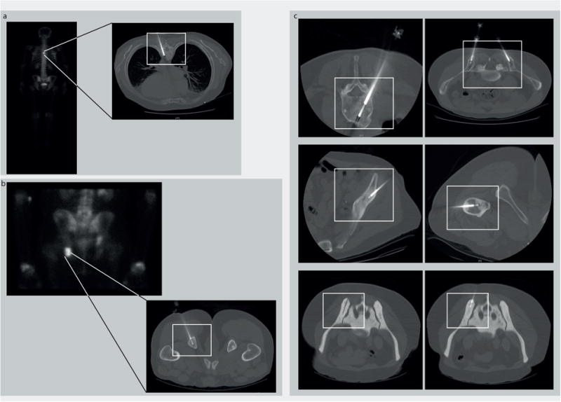
Figure 3.

Radiology images of different bone lesions in patients with metastatic prostate cancer who underwent a biopsy using the OnControl drill. (a) T6 vertebral body lesion and (b) lesion of the left ischial tuberosity. (Left) Scintigraphic image with clearly visible tracer uptake in both lesions. (Right) A corresponding computed tomography (CT) image demonstrating the intralesional biopsy needle. (c) CT images of biopsy procedures in multiple locations in several patients demonstrating safe use of the OnControl drill throughout the bony skeleton. CT-guided biopsy of an extremely dense bone lesion (lower panels: white rectangle, OnControl). The CT images confirm a technically successful biopsy.
