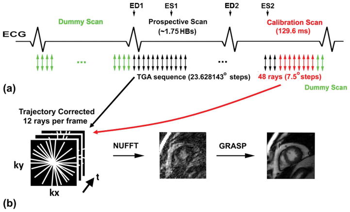
Figure 1.
(a) Pulse sequence diagram showing our sampling strategy. The first heartbeat was used to play pre-scan, dummy scans (green arrows) to approach steady state of magnetization. This is standard for all cine MRI with b-SSFP. Real-time, radial data were acquired in the subsequent 1.75 heartbeats using the TGA sequence (black arrows). Immediately after current data scan and before start of pre-scan, dummy scans for the following imaging plane, 48 rays of calibration data (red arrows; a pair of rays with opposite polarities per direction; 7.5° angular steps) were acquired in lieu of post-scan dummy scans (green arrows).

