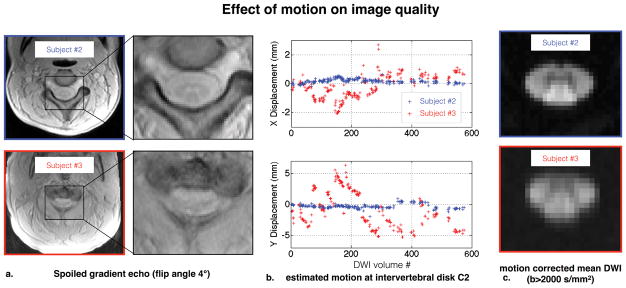
Figure 3.
Effect of the large motion of subject #3 on data quality (compared with subject #2 for illustration). a. Raw spoiled gradient echo images. Strong motion blurs the contrast between spinal cord gray and white matter. b. Lateral “X” (top) and antero-posterior “Y” (bottom) translations at C2, as estimated by the motion correction algorithm. Subject #3 showed abrupt motion with much larger amplitude than Subject #2 (~1cm in the Y direction). c. Motion corrected mean DWI (b>2000 s/mm2). Even with motion correction, Subject #3 presents more blurry boundaries suggesting worse data quality.

