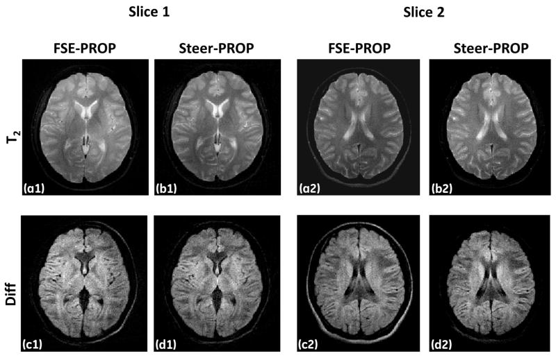
Figure 6.
Two slices of T2-weighted (a1, b1, a2, and b2) and diffusion-weighted (b = 750 s/mm2; c1, d1, c2, and d2) images from a human volunteer obtained at 1.5T using FSE-PROPELLER (first and third columns) and Steer-PROP (second and fourth columns) with TR = 4s, effective TE = 72ms, M = 8, N = 3 (for Steer-PROP), FOV = 24cm, slice thickness = 5mm, BW = ±62.5kHz, matrix size = 256×256, and NEX = 2.

