Figure 3.

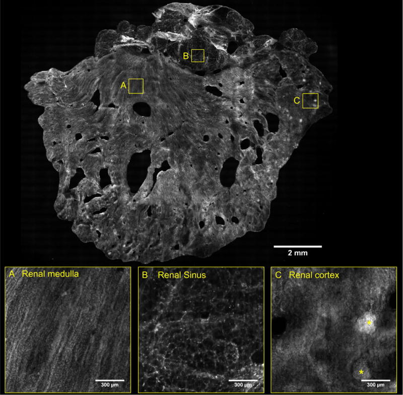
VR-SIM image of parenchymal margin from Case 7, with zooms of tubules and collecting ducts in the renal medulla (A), hilar adipose tissue in the renal sinus (B) and glomeruli (*) in the renal cortex (C).

VR-SIM image of parenchymal margin from Case 7, with zooms of tubules and collecting ducts in the renal medulla (A), hilar adipose tissue in the renal sinus (B) and glomeruli (*) in the renal cortex (C).