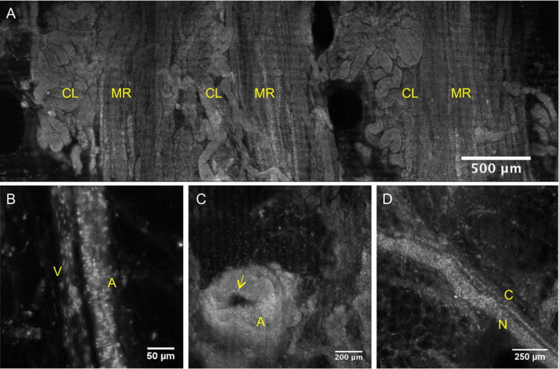
Figure 4.

VR-SIM images of tissue landmarks and structures commonly observed in the surgical margin images. A) Medullary rays (MR) marked by vertically ascending tubules and cortical labyrinth (CL) marked by intervening convoluted tubules. B) Arteriole (A) differentiated from an adjacent venule (V) by horizontally-oriented smooth muscle nuclei giving a ‘banded’ appearance and larger width, that the venule lacks. C) Artery (A) with a clear lumen and layers of wavy smooth muscle cell nuclei constituting the vessel wall, with apparent elastic lamina (yellow arrow) bordering the lumen. D) Peripheral nerve (N) and an associated small nutrient microvessel (C).
