Figure 2.
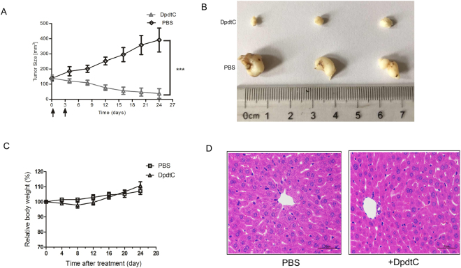
In vivo efficacy of DpdtC in the SK-OV-3 xenograft tumor model. (A) Mean tumor volumes of mice xenografted with SK-OV-3 cells and treated with DpdtC (5 mg/kg). There were 6 animals per treatment group. DpdtC treatment started as indicated in the graphs (black arrows). Error bars show ± SD. (***P < 0.001). (B) On day 24, xenograft tumor from each group were removed and photographed. Representative tumors in each group were shown. (C) Effect of DpdtC on nude mice body weight was determined using SK-OV-3 tumor-bearing nude mice. Mice were weighed at regular intervals during the whole period to monitor therapy-related toxicity. (D) Histological examination was conducted in nude mice post injection with DpdtC (5 mg/kg) for two times. Images (magnification, ×400) of liver from nude mice (n = 3) injected with PBS (−) or DpdtC (5 mg/kg) for two times were obtained by staining with hematoxylin and eosin. Scale bars, 50 μm.

