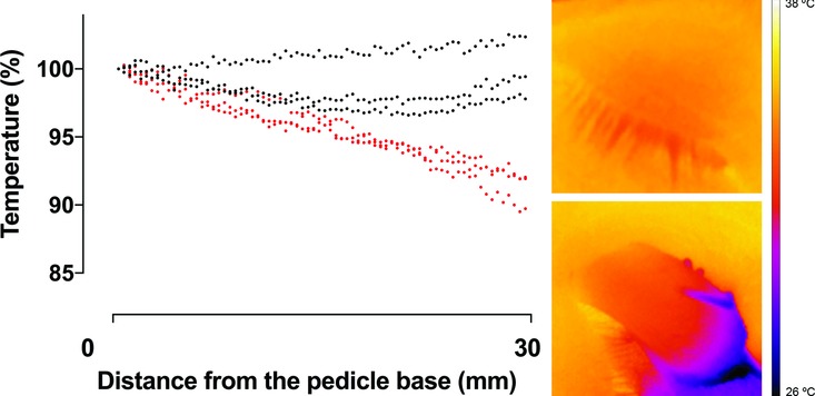
Figure 4.

Thermographic measurements showing a decrease in temperature from the pedicel base to the tip of the full-thickness eyelid flap (red symbols), compared with that of intact eyelids (black symbols), calculated as the percentage of the temperature in the pedicel base (n = 3). The images on the right are representative examples of thermographic images of an intact eyelid (top) and a dissected eyelid (bottom).
