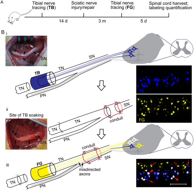
Figure 1.
Experimental paradigm (A) and schematic diagrams (B) showing sequential retrograde neuronal tracing. Sprague-Dawley rats, 3.5 months of age, were subjected to the first retrograde tracing by soaking the proximal stump of the lacerated left tibial nerve into 4% True Blue (TB) for 60 min to label the tibial motor neuron pool in the spinal cord (i). The tibial nerve stump was sutured to the distal one after application of the tracer. Fourteen days later, the left sciatic nerve was subjected to gap injury and subsequent repair with either a nerve autograft or an artificial nerve guidance conduit (ii). Sciatic nerve crush was performed as an additional control. Three months after the sciatic nerve injury and repair, the left tibial nerve was lacerated again but at 2 mm proximal to the previous laceration site, and the proximal stump of the tibial nerve was submerged into 5% FluoroGold (FG) for 60 min to label the spinal motor neurons that had regrew axons into the tibial nerve (iii). Five days later, rats were sacrificed for confocal microscopy to assess regeneration accuracy. The tibial motor neurons which faithfully re-innervated the tibial nerve were labeled by both TB and FG, showing white color (arrowheads), whereas those which failed to re-innervate the tibial nerve or did not regenerate axons at all were labeled by TB alone (filled arrows). The peroneal motor neurons whose axons were misdirected into the tibial nerve were labeled by FG alone (open arrows). SN, sciatic nerve; TN, tibial nerve; PN, peroneal nerve.

