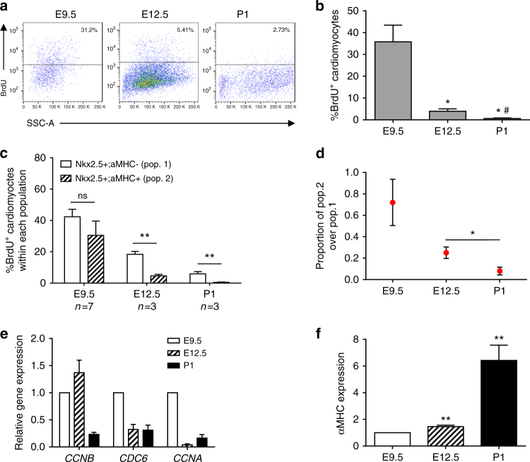
Fig. 4.
BrdU pulse-chase experiments substantiate decreasing proliferative capacity of CMs. a Representative flow cytometric analysis of BrdU incorporation. BrdU was given at E9.5, E12.5, or P1 αMHC-GFP mice 3 h prior to analysis. b Quantification of BrdU+ αMHC-GFP+ cells at E9.5, E12.5, and P1. (Student’s t test), *E12.5 or P1 vs. E9.5, #P1 vs. E12.5, p < 0.05. c Quantification of percent BrdU incorporation in Nkx2.5+;αMHC− and Nkx2.5+;αMHC+ cells at E9.5, E12.5, and P1. (Student’s t test), **p < 0.01. d Proportion of BrdU+ cells within the cardiomyocyte (Nkx2.5+/αMHC+) compared to progenitor (Nkx2.5+/αMHC−) populations at E9.5, E12.5, and P1. (Student’s t test), *p < 0.05. e qPCR analysis of αMHC-GFP+ cells reveals an age-dependent drop in expression of cell cycle genes (Ccna2, Ccnb1, Cdc6). f qPCR analysis of αMHC gene expression in αMHC-GFP+ cells from E9.5 to P1. All measurements shown are depicted as mean ± s.e.m

