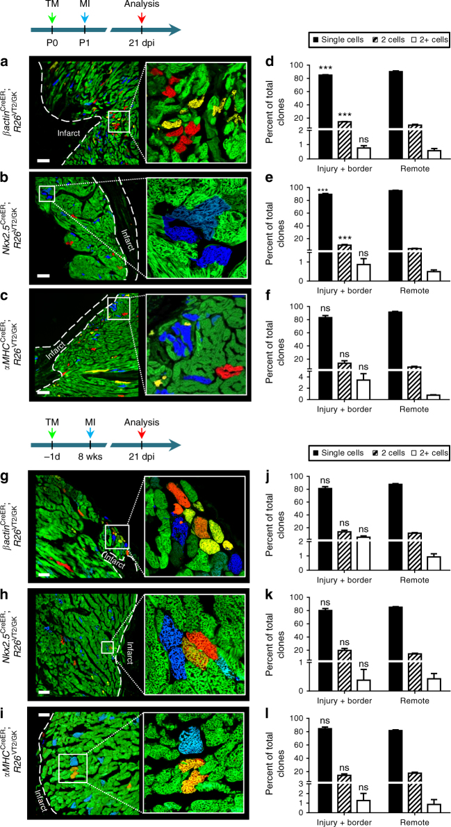
Fig. 6.
Myocardial injury activates CM proliferation in neonatal but not adult mice. Neonatal mice underwent left anterior descending artery (LAD) ligation at P1 and clonal analysis performed 21 days post injury. Representative confocal images of a βactinCreER; R26VT2/GK, b Nkx2.5CreER; R26VT2/GK and c αMHCCreER; R26VT2/GK heart (left ventricle) sections. Insets show close-up of boxed regions. d–f Quantification of clonal formation following neonatal injury. LAD ligation was performed in 8-week-old mice (g, h, i), followed by clonal analysis 3 weeks post-injury. Representative fluorescent microscope images of the infarct and border zone of g βactinCreER; R26VT2/GK, h Nkx2.5CreER; R26VT2/GK and i αMHCCreER; R26VT2/GK hearts (left ventricle). j–l Quantification of clonal formation following adult injury. White dashed line marks infarct area. (Tukey’s multiple comparison test), ***p < 0.001 to remote. All measurements shown are depicted as mean ± s.e.m

