Figure 1.
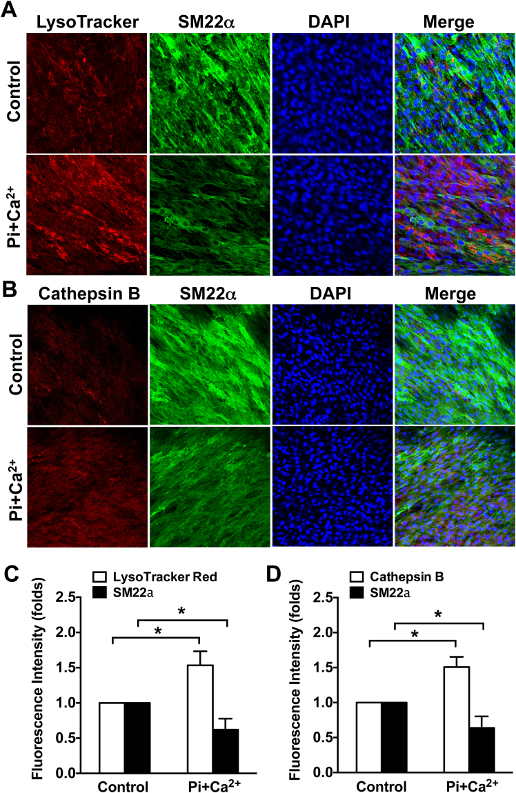
Lysosomal function is increased in calcifying VSMCs. Confluent rat aortic VSMCs were cultured on a chamber slide in a Pi + Ca2+ (3.5 mM phosphate/3 mM calcium) calcification medium for 7 days, the function of lysosome was evaluated by the LysoTracker red and cathepsin B magic red staining. (A and C) Representative immunofluorescent staining images and quantitative results showing increased LysoTracker intensity and decreased SM22α expression in calcifying VSMCs. (C–D) Immunofluorescent staining images and quantitative results showing effects of calcification medium on cathepsin B activity and SM22α expression. The fluorescent intensity of lysosomes was analyzed using Image J software. At least 5 fields per condition and 3 independent experiments were performed. All Values are presented as mean ± SD. *P < 0.05.

